Description of an Athlete’s foot
Athlete’s foot (tinea pedis) is a common, persistent infection of the foot caused by a dermatophyte, a microscopic fungus that lives on dead tissue of the hair, toenails, and outer skin layers. These fungi thrive in warm, moist environments such as shoes, stockings, and the floors of public showers, locker rooms, and swimming pools. Athlete’s foot is transmitted through contact with a cut or abrasion on the plantar surface (bottom) of the foot. In rare cases, the fungus is transmitted from infected animals to humans. Dermatophyte (skin) infections cause raised, circular pimples or blisters that resemble the lesions caused by ringworm. The infections are named after the part of the body they infect. Tinea pedis, therefore, refers to an infection of the feet.
Incidence and prevalence of athlete's foot
Athlete’s foot is most common in men from the teenage years to the early 50s. Prevalence is affected by personal hygiene and daily activity. People with compromised immune systems are at greater risk.
Causes of an Athlete’s foot
There are at least four dermatophytes that can cause tineas pedis. The most common is trichophyton rubrum.
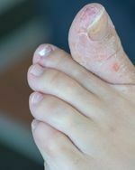
There are four common forms of athlete’s foot. The most common is an annoying, persistent itching of the skin on the sole of the foot or between the toes (often the fourth and fifth toes). As the infection progresses, the skin grows soft. The center of the infection is inflamed and sensitive to the touch. Gradually, the edges of the infected area become milky white and the skin begins to peel. There may also be a slight watery discharge.
In the ulcerative type, the peeling skin becomes worse. Large cracks develop in the skin, making the patient susceptible to secondary bacterial infections. The infection can be transmitted to other parts of the body by scratching, or contamination of clothing or bedding.
The third type of tinea infection is often called “moccasin foot.” In this type, a red rash spreads across the lower portion of the foot in the pattern of a moccasin. The skin in this region gradually becomes dense, white, and scaly.
The fourth form of tinea pedis is inflammatory or vesicular, in which a series of raised bumps or ridges develops under the skin on the bottom of the foot, typically in the region of the metatarsal heads. Itching is intense and there is less peeling of the skin.
People with acute tinea infections may develop similar outbreaks on their hands, typically on the palms. This trichophyde reaction, also known as tineas manuum, is an immune system response to fungal antigens (antibodies that fight the fungal infection).
Diagnosis of an Athlete’s foot
Diagnosis is made by visual observation of the symptoms. The podiatrist eliminates the possibility of a bacterial infection by performing a microscopic examination of skin scrapings to determine the type of fungus causing the infection. Other tests include growing a fungal culture from skin scrapings and examining the patient’s foot under an ultraviolet light.
Treatment of an Athlete’s foot
Tinea infections may disappear spontaneously or persist for years. They are difficult to eliminate and often recur. Best results usually are obtained with early treatment before the fungal infection establishes itself firmly. Antifungal drugs may be used to fight the infection.
In most cases, 4 to 6 weeks of treatment clears up the infection. If the infection becomes systemic, stronger antifungal medication may be prescribed.
If the infection is bacterial, a course of oral antibiotics may be prescribed.
The information provided in this web site is not intended to be a substitute for medical examination, diagnosis or treatment. The material is provided for information purposes alone.